Inside Preclinical Ocular Research: With Dr. Simone Iwabe, DVM, PhD, DACVO, and Randy Wheeland, BS

Altasciences has been at the forefront of ophthalmic drug development for over 30 years, having completed more than 100 ocular studies to assess the safety of new drug products intended for human use.
To uncover what sets Altasciences’ early-phase ophthalmic solutions apart from other CROs, we spoke with Dr. Simone Iwabe, DVM, Director of Veterinary Services, and Randy Wheeland, BS, Pathology Associate III. Here’s what they had to say:
Q. What specialized expertise is required to support early-phase preclinical ocular studies effectively?
SI: Supporting preclinical ocular studies requires a high level of technical proficiency and attention to detail. Here at Altasciences, our expertise includes topical dosing, capturing images using a fundus camera, optical coherence tomography (OCT), confocal microscopy, and electroretinography to measure retinal activity. Whether conducting tonometry or measuring corneal thickness, our technicians have received the specialized training required to carry out these tasks with precision.
Q. How do your teams work together on the advanced ocular studies you conduct?
SI: The teams across our four preclinical sites follow fully harmonized and integrated procedures. In addition, our study directors have the expertise needed to oversee and conduct all ocular studies.
Our necropsy team consists of experts skilled in collecting and analyzing eye tissues.
Q. What instruments do you use to conduct ocular studies?
SI: We use two fundus cameras to capture color images of the back of the eye and take photographs before, during, and after the study. This is especially useful for monitoring intravitreal injections, whether suspensions or inserts, as these images help track the test article’s progression over time.
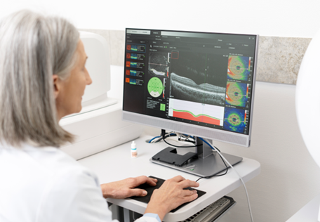
Another frequently used device is the Heidelberg Spectralis HRA/OCT, which is essential for observing retinal health throughout the study. The Spectralis provides high-resolution black-and-white images of the fundus. We can place a green line on the machine to mark the OCT area, which gives us an in vivo cross-section of the retina, ensuring it remains normal throughout the study.

We recently incorporated confocal microscopy to our capabilities to analyze changes in the cornea, and pupillometry to evaluate changes in the pupil response for more comprehensive analyses of ocular health and test article impact.
Q. What are some necropsy techniques that can be used for ocular studies?
RW: In necropsy we can analyze the entire eye as a single block or dissect it into various portions, which is vital for comprehensive ocular studies, and something not all CROs can do. We remove the eye for dissection, including the iris, and the ciliary body, and can even separate the retina from the choroid, which is uncommon in many facilities. Additionally, we can freeze these issues in liquid nitrogen and send them for further study.
We tailor our approach based on our clients’ needs, focusing on specific areas related to the test article. For example, if a sponsor is interested in understanding how the test article affects the meniscus tissue, we can isolate and process that tissue for in-depth analysis and identify any irregularities.
Q. What ocular procedures can Altasciences perform in preclinical studies?
SI: Our procedures span from minor interventions to highly complex surgeries; we conduct both routine and cutting-edge studies.
We’re fully specialized and equipped to perform a variety of ocular procedures, such as subretinal, intravitreal (IVT), suprachoroidal, sub-Tenon, intracameral, intralacrimal, and subconjunctival injections—with the capability to perform invasive anterior and posterior segment surgeries using microscopes and a Stellaris machine. This allows us to conduct cataract surgeries, vitrectomies, or semi-automated subretinal injections.
We also have the expertise to develop ocular animal models tailored for efficacy studies, and are fully prepared to accommodate any additional requirements from the sponsor.
In Conclusion
From comprehensive ocular assessments to complex surgical interventions, we have the knowledge, experience, and equipment to handle the full spectrum of your ophthalmic research needs.
We are here to support your next project. If you’re ready to discuss your ophthalmic study requirements or explore opportunities for preclinical drug development in your pipeline, don’t hesitate to contact us.
Learn more about our comprehensive ophthalmic drug development services.
This article was posted in April 2025, and edited in May 2025.